
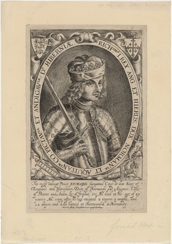
Richard’s death unfolded over days, leaving room for a different question from Caesar’s: what happens after the initial wound has been survived?
A wound in the Limousin
Richard I was wounded by a crossbow bolt while besieging Châlus in 1199. He died on 6 April. A modern examination of his preserved heart summarises a wound near the left shoulder and considers gangrene or septicaemia a reasonable explanation for the subsequent death.[1]
That study investigated the embalming of the heart. It did not isolate a pathogen from the fatal wound or perform a modern autopsy of the whole body. The distinction matters: an infected wound is a plausible interpretation of the course, while a named bacterium, toxin or precise sepsis syndrome would exceed the evidence.[1]
The familiar stories about the archer and royal forgiveness belong to the wider chronicle tradition. This case keeps its attention on the documented wound, delayed death and the clinical problem of deteriorating tissue.
The injury after the impact
A penetrating wound may carry foreign material into tissue and leave areas without a blood supply. Assess its extent, contamination and signs of infection. Modern wound care removes dirt and foreign material and addresses dead (necrotic) tissue, with appropriate expertise.[2]
Tetanus prevention requires its own assessment of the wound and immunisation history. Antibiotics used for a wound infection do not replace indicated tetanus vaccination or immunoglobulin. The historical record here does not establish tetanus as Richard’s disease.[2]
A surface dressing can conceal the course beneath it. Increasing illness after a wound requires renewed examination and escalation. The passage of several days is no guarantee that the danger has passed.
After the wound is dressed
Follow the problems that can remain after the initial injury has been survived.
Material in the wound
Wound assessment considers contamination, retained material and injured tissue. Appropriate cleaning and removal require a safe clinical approach.[2]
Devitalised tissue
Tissue that has lost viability can require debridement. A dressing cannot establish that an underlying source is controlled.[2][3]
Possible organ dysfunction
Infection accompanied by systemic deterioration requires urgent assessment for sepsis and other causes.[3]
Material in the wound
Wound assessment considers contamination, retained material and injured tissue. Appropriate cleaning and removal require a safe clinical approach.[2]
Devitalised tissue
Tissue that has lost viability can require debridement. A dressing cannot establish that an underlying source is controlled.[2][3]
Possible organ dysfunction
Infection accompanied by systemic deterioration requires urgent assessment for sepsis and other causes.[3]
Educational mechanism study. These panels explain the case and are not standalone treatment protocols.
When infection affects the whole patient
Sepsis is life-threatening organ dysfunction caused by a dysregulated response to infection. Modern care evaluates the entire patient and the likely source. Deterioration in mental state, circulation, breathing or renal function changes the urgency of assessment and treatment.[3]
Antimicrobial treatment is selected for the suspected source and local guidance, with urgency determined by clinical risk. Investigation and reassessment continue alongside treatment. A single wound description cannot determine the antibiotic required.[4]
Source control may require drainage or removal of infected and devitalised tissue. The Surviving Sepsis Campaign advises identifying an anatomical focus requiring such intervention and acting as soon as medically and logistically practical. Antimicrobial therapy does not make that assessment redundant.[3]
An earlier opportunity
Modern care might alter the course at several points: initial wound assessment, management of contamination, tetanus protection where indicated, recognition of deterioration, antimicrobial treatment and surgery for an infected focus. The relevant opportunity extends beyond the moment the bolt is removed.[2][3][4]
That broader chain of care makes Richard a useful companion to the ancient cases. Immediate haemorrhage and later infection threaten a casualty on different timescales. Surviving the first does not complete the treatment of the wound.
Could Richard have survived?
A contaminated wound with subsequent infection presents several opportunities for effective modern treatment. That makes a changed outcome plausible. The missing anatomy and microbiology prevent certainty about Richard himself, or about which intervention would have been decisive.[1][2][3]
At the bedside
Three decisions about the clinical problem. The supplied findings are teaching scenarios; they are not additional historical observations.
Sources and notes
Sources checked 10 October 2026. Historical testimony, mechanism research and clinical guidance are identified separately. Modern guidance retains its stated setting and population.
- Charlier et al., The embalmed heart of Richard the LionheartPrimary researchScientific Reports 3, 1296, 2013. Historical injury summary and biological analysis of embalming; cause of death remains inferred.
- CDC, Wound management to prevent tetanusClinical guidanceReviewed 8 September 2026. US guidance; wound care and separate tetanus prevention. No US vaccine schedule is reproduced.
- Surviving Sepsis Campaign, international guidelinesClinical guidance2021. Adults in hospital; definition, source control and reassessment.
- NICE NG253, Suspected sepsis in people aged 16 or overClinical guidance19 November 2025. UK recognition and management; excludes pregnancy and recent pregnancy. Antimicrobial choice follows source and local policy.
Artwork and provenance
Renold Elstrack, Richard I of England, published 1618. National Gallery of Art, Washington. Rosenwald Collection
A seventeenth-century imagined portrait of Richard I, published more than four centuries after his death. Images are displayed in monochrome and framed to fit the page. The source images are served from this website.
Public domain / National Gallery of Art Open Access. Museum or collection record · Rights record · Original image