

ECG interpretation.
An adult prehospital course in 12-lead reading, rhythm reasoning and clear clinical handover.
Shock or no shock?
Watch the heart and ECG together. Choose an action and see the response.
Start the free challengeNo enrolment required.
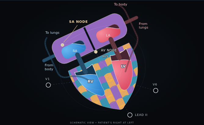 Explore the heart in motion
The irregular rhythm.
Count the ventricular events, compare rhythms and inspect a clinical recording. Five checks explain the reasoning behind each answer.
Try the free lessonNo registration · progress saved on this device
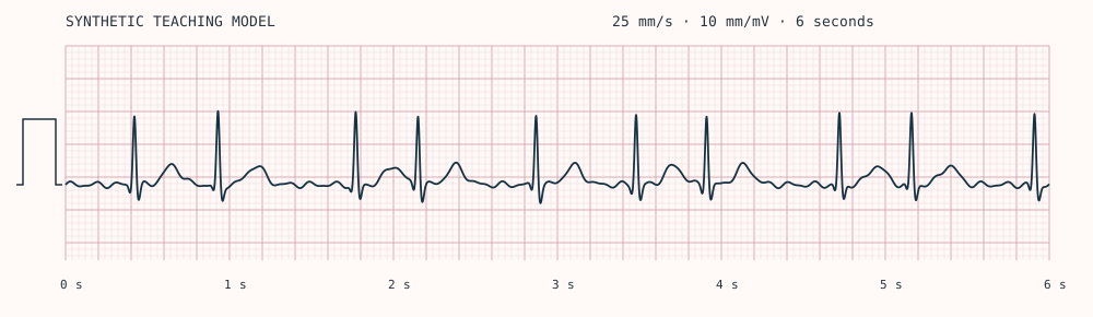
The full ECG course.
Ten lessons in rhythm and 12-lead interpretation, with 65 decision checks and a 20-question final assessment.
Study 18 rhythm diagrams alongside 13 clinical ECG recordings, with conduction anatomy, enlarged views and reveal practice.
Price on application. Enrolment is currently closed.
Already have access?
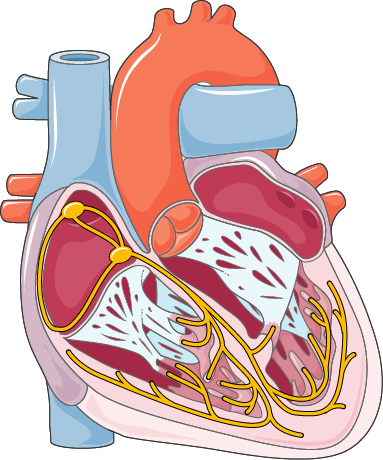
Image source
Servier Medical Art, Heart conduction · CC BY 4.0. Original image, scaled for display.
View the ten-module syllabus
- IConduction, paper and the normal beatSA node to Purkinje fibres, P–QRS–T and calibration
- IIReading an ECG, step by stepRate, rhythm, intervals, lead views and a consistent read
- IIIShockable and non-shockableVF, pulseless VT, PEA and asystole
- IVCommon rhythms and arrhythmiasSinus rhythms, AF, flutter, SVT and ectopy
- VHeart blocks and broad-complex rhythmsAV blocks, VT and the patient’s condition
- VIIschaemia in contextChange, comparison and uncertainty
- VIISubtle high-risk patternsFindings that need a second look
- VIIIMimics and metabolic cluesAlternative explanations for the trace
- IXThe first ten minutesReassessment and clear communication
- XInterpretation under pressureUnfamiliar cases and a structured handover
Original rhythm diagrams are labelled as synthetic teaching models. Clinical recordings retain their source credits and ECG-paper grids. A tracing is one part of the clinical assessment.